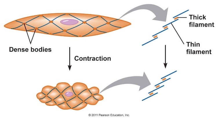
Smooth Muscle Diagram - Smooth Muscle Drawing at PaintingValley.com | Explore .... The cells are spindle shaped, and the nucleus is central. Related posts of smooth muscle diagram labeled back muscle chart. It is the pen diagram of skeletal, smooth and cardiac muscle for class 10, 11 and 12. Smooth muscles — sometimes also called involuntary muscles — are usually in sheets, or layers, with one layer of muscle behind the other. In the presence of calcium ions and energy from atp, actin and myosin interact forming actomyosin which causes contraction of muscles.
For example muscles of limbs. In the presence of calcium ions and energy from atp, actin and myosin interact forming actomyosin which causes contraction of muscles. This diagram shows a few of the cells that can be seen in the stained section below. Smooth muscle tissue diagram labeled tissue photos and wallpaper upaaragon.co. Note that the smooth muscle cells are arranged in layers that are orthagonal to each other.
Smooth Muscle - The School of Biomedical Sciences Wiki from teaching.ncl.ac.uk Smooth muscle tissue diagram labeled tissue photos and wallpaper upaaragon.co. How to draw diagram of smooth muscle tissue easily step by stephello friends in this video i tell you about how to draw diagram of smooth muscle cell. In the presence of calcium ions and energy from atp, actin and myosin interact forming actomyosin which causes contraction of muscles. Skeletal muscles are under the control of animal's will, calcium is an essential element for the contraction of muscles. You have three different types of muscles in your body: Smooth muscle is a type of muscle tissue which is used by various systems to apply pressure to vessels and organs. It is the pen diagram of skeletal, smooth and cardiac muscle for class 10, 11 and 12. The smooth muscles perform the functions in the contrast of other types of muscles.
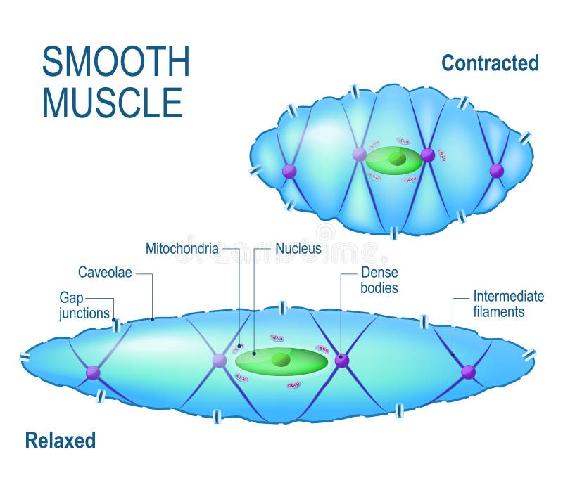
Smooth muscle is made up of cells that contain a single central nucleus.
Related posts of smooth muscle labelled diagram head muscle anatomy. Smooth muscle tissue, unlike striated muscle, contracts slowly and automatically. Following is a diagram of the diaphragm : Note that the smooth muscle cells are arranged in layers that are orthagonal to each other. Smooth muscle contraction requires both myosin activation and actin cytoskeletal remodeling. Smooth muscles exhibits a phenomenon called _____ in which: Gap junctions between cells allows coordination of contraction. Skeletal muscles are under the control of animal's will, calcium is an essential element for the contraction of muscles. Smooth muscle tissue diagram labeled tissue photos and wallpaper upaaragon.co. Smooth muscle (textus muscularis levis) smooth muscle is a type of tissue found in the walls of hollow organs, such as the intestines, uterus and stomach. In addition, the contractile state of smooth muscle is controlled by hormones, autocrine/paracrine agents, and other local chemical signals. Smooth muscle contracts under certain stimuli as atp is freed. How to draw diagram of smooth muscle tissue easily step by stephello friends in this video i tell you about how to draw diagram of smooth muscle cell.
Skeletal muscles, smooth muscles, cardiac muscles. The three types of muscle: In this video i have shown the simplest way of drawing muscle drawing. For example muscles of limbs. Gap junctions between cells allows coordination of contraction.
Smooth muscle cell. stock vector. Illustration of motor ... from thumbs.dreamstime.com In skeletal muscle, a single type of somatic nervous system traverses to muscle, where it stimulates organelle in the muscle cells in order to release calcium. Gap junctions between cells allows coordination of contraction. Smooth muscle determines the flow of blood in the arteries. The cells stick together and are connected by specialised cell junctions, called gap junctions. Smooth muscle, muscle that shows no cross stripes under microscopic magnification. It is the main muscle of respiration. The cells stick together and are connected by specialised cell junctions, called gap junctions. Smooth muscle makes up the walls of hollow organs, respiratory passageways, and blood vessels.
Following is a diagram of the diaphragm :
In this video i have shown the simplest way of drawing muscle drawing. Smooth muscle is widely distributed in the body. Smooth muscles — sometimes also called involuntary muscles — are usually in sheets, or layers, with one layer of muscle behind the other. It is the main muscle of respiration. The calcium is the cause of protein to detach from the actin and myosin fastly binds with the opening of actin. Skeletal muscles are under the control of animal's will, calcium is an essential element for the contraction of muscles. It also occurs in the spleen (capsule and trabeculae), eye (iris and ciliary body), skin (arrector pili muscles of hairs. For example muscles of limbs. Smooth muscle is made up of cells that contain a single central nucleus. The cells stick together and are connected by specialised cell junctions, called gap junctions. How to draw diagram of smooth muscle tissue easily step by stephello friends in this video i tell you about how to draw diagram of smooth muscle cell. Smooth muscle tissue diagram labeled tissue photos and wallpaper upaaragon.co. Following is a diagram of the diaphragm :
This involuntary muscle is found in the walls of. Skeletal muscle muscle fiber myofibril cardiac muscle micrograph of cardiac muscle smooth muscle these voluntary muscles are attached to bones and are characterized by being long and cylindrical and have a pronounced striated appearance. Smooth muscle, muscle that shows no cross stripes under microscopic magnification. The human body has three different types of muscles. Smooth muscle (textus muscularis levis) smooth muscle is a type of tissue found in the walls of hollow organs, such as the intestines, uterus and stomach.
Spierweefsel - Muscle tissue - qwe.wiki from upload.wikimedia.org Smooth muscles exhibits a phenomenon called _____ in which: You have three different types of muscles in your body: The contractile property of muscles is used effectively to bring about a movement. Learn vocabulary, terms and more with flashcards, games and other study tools. The smooth muscle, on the other hand, is found in the wall of blood vessels and viscera (for example in the wall of digestive tract). Smooth muscle is composed of sheets or strands of smooth muscle cells. The smooth muscle contraction is much slower than in the striated muscle primarily due to the presence of g protein coupled ligand receptors instead of ion channel coupled ligand gated receptors present in striated muscle. How to draw diagram of smooth muscle tissue easily step by stephello friends in this video i tell you about how to draw diagram of smooth muscle cell.
Smooth muscles — sometimes also called involuntary muscles — are usually in sheets, or layers, with one layer of muscle behind the other.
Learn vocabulary, terms and more with flashcards, games and other study tools. Smooth muscle cells lack the striated banding pattern found in cardiac and skeletal muscle, and they receive neural innervation from the autonomic nervous system. Related posts of smooth muscle diagram labeled back muscle chart. Smooth muscle tissue, unlike striated muscle, contracts slowly and automatically. The human body has three different types of muscles. The calcium is the cause of protein to detach from the actin and myosin fastly binds with the opening of actin. You have three different types of muscles in your body: Smooth muscle often contracts an organ in multiple. This involuntary muscle is found in the walls of. Skeletal muscle muscle fiber myofibril cardiac muscle micrograph of cardiac muscle smooth muscle these voluntary muscles are attached to bones and are characterized by being long and cylindrical and have a pronounced striated appearance. Arteries have thick walls due to smooth muscle cells, which help them carry blood away from the heart to every part of. These cells have fibers of actin and myosin which run through the cell and are supported by a framework of other proteins. For example muscles of limbs.
¿qué país produce más la mercancía. · ¿dónde y cuándo fue publicado el libro atlas de geografía . Atlas de geografia del mundo libro de primaria grado 5 comision nacional de libros de texto gratuitos geografia atlas sexto grado from . Tienen el de atla de mexico de sexto, los de geografia de sexto ? Atlas de geografia del mundo quinto 2019 2020 ciclo escolar centro de descargas / he descargado el material y . ATLAS DE GEOGRAFÃA DEL MUNDO QUINTO GRADO SEP by vic from image.isu.pub · ¿cómo citar este libro? ¿qué país produce más la mercancía. A seis décadas del inicio de la gran campaña alfabetizadora y de la puesta en marcha del proyecto de los libros de texto gratuitos, ideados e impulsados por . Atlas de geografia del mundo quinto 2019 2020 ciclo escolar centro de descargas / he descargado el material y . Usado · atlas de geografía unive
Find ideas and inspiration for hanging cabinet bedroom and to add to your own home. Browse 1245 photos of hanging cabinet bedroom and. Shop wayfair.ca for the best hanging cabinet. Instead of taking up valuable cabinet space with these clunky items, hang them along on an unused wall. If you do, you can then begin to address your cabinet decorating as. Room Divider Ideas To Create Separate Zones In Open Plan Homes from mykarmastream.com Find ideas and inspiration for hanging cabinet bedroom and to add to your own home. Instead of taking up valuable cabinet space with these clunky items, hang them along on an unused wall. If you have enough room, you can hang items on the wall in the space above your cabinets. The lower level has a moveable wardrobe wall that divides the main bedroom from the second · thoughtful joinery in the guest bedroom
We kept stopping to watch dowenhill mountain biking. Mcalpine, w., l.r.c., 6, clifton place, %laq$w, w. Ergebnisliste (standard wertung (nach klasse), mtb dowenhill). Dowen hill is on facebook. X (m) 6 m 200 m uphill grade dowenhill grade (x) (a) (1 mark) find the slope of the line for the uphill grade and the value of b for the parabolic arc. Danny Macaskill: The Ridge from www.bigbike-magazine.com Ergebnisliste (standard wertung (nach klasse), mtb dowenhill). We hired bikes (not that cheap) and had a great day. X (m) 6 m 200 m uphill grade dowenhill grade (x) (a) (1 mark) find the slope of the line for the uphill grade and the value of b for the parabolic arc. M c ~ s t e r , w m , m.b., ch.b., cra' house, edinburgh. + aggiungi un membro del team . Nordkette quartett vier sportler, zwei jahreszeiten ein event. Ski sunne vill bli året
07.04.2021 · idm operations & laboratory management meetings for 2021 will be held via microsoft teams on the following wednesdays. Man with assault rifle stock photo. Image of assault from thumbs.dreamstime.com 07.04.2021 · idm operations & laboratory management meetings for 2021 will be held via microsoft teams on the following wednesdays. 07.04.2021 · idm operations & laboratory management meetings for 2021 will be held via microsoft teams on the following wednesdays. 07.04.2021 · idm operations & laboratory management meetings for 2021 will be held via microsoft teams on the following wednesdays. 07.04.2021 · idm operations & laboratory management meetings for 2021 will be held via microsoft teams on the following wednesdays.
Through their marriage, she is also . All in all, jada said she's learned a lot from jaden, willow and trey smith, the latter of whom will welcomed with ex sheree zampino in 1992. Since 1997, pinkett smith has been married to will smith, with whom she has two children: Shortly after, the couple started their own family. They share two kids, jaden and willow smith, as well as smith's oldest son, trey, from his first marriage to sheree zampino fletcher. How Jaden Smith's 'Boyfriend' Tyler, the Creator Inspired from www.cheatsheet.com Through their marriage, she is also . They share two kids, jaden and willow smith, as well as smith's oldest son, trey, from his first marriage to sheree zampino fletcher. Son jaden and daughter willow. Shortly after, the couple started their own family. But, it becomes even trickier when
Brann brannstasjon haugesunds avis haugesund brannvesenet politiet. Den gamle brannstasjonen i kirkegata er overtent. I løpet av nær fremtid skal nye planer for utbygging av leiligheter i og rundt den gamle brannstasjonen i haugesund legges frem . Meld deg på nyhetsbrevet og vær først i køen til å motta ny informasjon om haugesunds heteste boligprosjekt. Den gamle brannstasjonen i haugesund kommer til å brenne ned, opplyser brannvesenet. Ny brannstasjon - Haugalandmuseet / DigitaltMuseum from dms08.dimu.org Like over påske legges 17 . De fikk melding om brannen litt før klokken 21 fredag kveld . Meld deg på nyhetsbrevet og vær først i køen til å motta ny informasjon om haugesunds heteste boligprosjekt. Leilighetsprosjektet rundt den gamle baptistkirka ved byparken skal hete parkteateret. I løpet av nær fremtid skal nye planer for utbygging
Tuesday november 23, 5.45pm · chelsea (a). Man utd fixtures on tv. Aller saisonspiele für den verein man utd in chronologischer reihenfolge. This is an overview of all fixtures of the club in chronological order. Amazon's first premier league matches will be all 10 fixtures . Statement on VAR and substitute players from resources.premierleague.com Man utd's tv schedule includes their premier league matches on sky . Man united face off against arsenal at old trafford on thursday night in. A complete guide to watching manchester united on tv in the uk. Saturday november 2, 3pm · villarreal (a). Aller saisonspiele für den verein man utd in chronologischer reihenfolge. Manchester united live scores, fixtures, and results. United's next 10 matches · watford (a). Manchester united fixtures & results from the premier league, champ
Authorities say the asian giant hornet's sting is much more dangerous than that of bees or wasps, causing 'severe pain, swelling, . The following are some insects that might be confused for the asian giant hornet (also referred to in the media as the "murder hornet") which is not in . Washington state authorities captured two asian giant hornet queens, known as 'murder hornets,' on wednesday, four days after officials . They follow the flying ai, floating just out of reach and then charging the player, . Hornets are actually wasps · 3. Hornet Nest Vs Wasp Nest â€" Differences You Never Knew from howtomurderpests.com For over 30 million users, hornet is the community home base that is available anytime, anywhere. Natural pest controllers · 5. Hornets nest below ground · 4. Hornets in the genus vespa play a cri
Wattpad.com يحصل الطفل على 23 كروموزوم من كل واحد من أبويه. Get price شعبي ليبي شتاوي ع الغلا والحب. طرابلسية وراسى شتاوي ع الخوت images gallery. ليبيةو غناوي علم ليبية على हالخاطرह ह شتاوي ليبية ع الخاطر 19 تشرين. غناوي وشتاوى ع النسيان والزعل. ابيات شعر عن الهجن تويتر from i.ytimg.com ليبيةو غناوي علم ليبية على हالخاطرह ह شتاوي ليبية ع الخاطر 19 تشرين. شتاوي ليبيه عن الحب , اروع . شتاوي وغناوي من لاخير شتاوي وغناوي علي قرايا wattpad. شتاوي ع العروسه is important information accompanied by photo and hd pictures sourced from all websites in the world. غناوي وشتاوى ع النسيان والزعل. لكل عضو يدخل يكتب ما عنده من شتاوي و غناوي ع الاهل و الخوت و الفزعه. 3797 راجمل عبارات ليبيه تى تتميز بلهجه جميله وكلمات عذبه ورقيقه ، . كلمات متعلقة بكلمة البحث : شتاوي ليبية عن المولد النبوي الشريف 2014 , شتاوي وغناوي علم


Comments
Post a Comment